- Automatic segmentation saves time
-
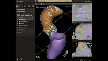
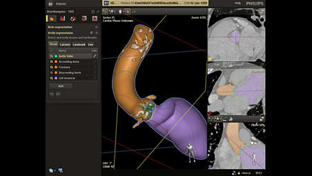
Automatic segmentation saves time
Once you load a pre-operative DICOM compliant CT dataset and a 3D volume is rendered, HeartNavigator automatically segments tissue, anatomical structures, landmarks, calcium, and planes of the heart for TAVR/TAVI. To facilitate mitral valve replacement, left atrial appendage closure (LAAC) and other procedures, HeartNavigator automatically segments the entire heart. - Automatic landmarks to stay on track
-
Automatic landmarks to stay on track
For additional guidance, HeartNavigator automatically places landmarks on a large variety of anatomical structures, including the ostia of the coronaries, 3 nadirs, etc. They can be manually adapted as needed. - Automatic measurements may help streamline your planning
-
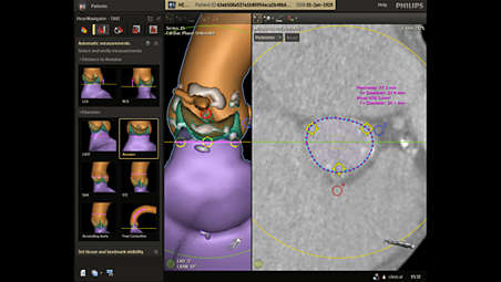
Automatic measurements may help streamline your planning
A single click creates area, perimeter, diameter and distance measurements of anatomical structures for TAVR or TAVI procedures. The measurements are performed within the detected anatomical planes and are shown on the displayed centerline. For all other SHD procedures, manual measurements are provided. - Calcification visualization to avoid potential complications
-
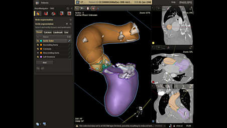
Calcification visualization to avoid potential complications
The software enhances insight into the distribution of calcifications in the ascending aorta, aortic valve annulus, and left ventricle. By determining the severity and location of calcification, you can avoid potential complications during procedures. - Automatic view planning aids positioning
-
Automatic view planning aids positioning
HeartNavigator automatically determines the optimal projection angles to use during the procedure. This can avoid the need to acquire multiple aortograms. Projections can be recalled by the tableside for further savings. - Integrated live image guidance supports precise navigation
-
Integrated live image guidance supports precise navigation
You can get real-time feedback to guide navigation through the vasculature. The 3D CT volume rendering of the ascending aorta can be matched with live fluoro to show the exact position of catheters and devices in relation to the reference image. The 3D CT rendering moves in sync with the system's movements during the procedure.
Automatic segmentation saves time
Automatic segmentation saves time
Automatic segmentation saves time
Automatic landmarks to stay on track
Automatic landmarks to stay on track
Automatic landmarks to stay on track
Automatic measurements may help streamline your planning
Automatic measurements may help streamline your planning
Automatic measurements may help streamline your planning
Calcification visualization to avoid potential complications
Calcification visualization to avoid potential complications
Calcification visualization to avoid potential complications
Automatic view planning aids positioning
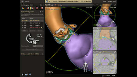
Automatic view planning aids positioning
Automatic view planning aids positioning
Integrated live image guidance supports precise navigation
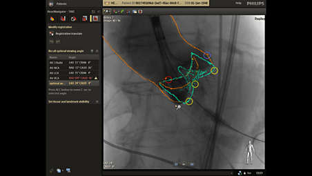
Integrated live image guidance supports precise navigation
Integrated live image guidance supports precise navigation
- Automatic segmentation saves time
- Automatic landmarks to stay on track
- Automatic measurements may help streamline your planning
- Calcification visualization to avoid potential complications
- Automatic segmentation saves time
-
Automatic segmentation saves time
Once you load a pre-operative DICOM compliant CT dataset and a 3D volume is rendered, HeartNavigator automatically segments tissue, anatomical structures, landmarks, calcium, and planes of the heart for TAVR/TAVI. To facilitate mitral valve replacement, left atrial appendage closure (LAAC) and other procedures, HeartNavigator automatically segments the entire heart. - Automatic landmarks to stay on track
-
Automatic landmarks to stay on track
For additional guidance, HeartNavigator automatically places landmarks on a large variety of anatomical structures, including the ostia of the coronaries, 3 nadirs, etc. They can be manually adapted as needed. - Automatic measurements may help streamline your planning
-
Automatic measurements may help streamline your planning
A single click creates area, perimeter, diameter and distance measurements of anatomical structures for TAVR or TAVI procedures. The measurements are performed within the detected anatomical planes and are shown on the displayed centerline. For all other SHD procedures, manual measurements are provided. - Calcification visualization to avoid potential complications
-
Calcification visualization to avoid potential complications
The software enhances insight into the distribution of calcifications in the ascending aorta, aortic valve annulus, and left ventricle. By determining the severity and location of calcification, you can avoid potential complications during procedures. - Automatic view planning aids positioning
-
Automatic view planning aids positioning
HeartNavigator automatically determines the optimal projection angles to use during the procedure. This can avoid the need to acquire multiple aortograms. Projections can be recalled by the tableside for further savings. - Integrated live image guidance supports precise navigation
-
Integrated live image guidance supports precise navigation
You can get real-time feedback to guide navigation through the vasculature. The 3D CT volume rendering of the ascending aorta can be matched with live fluoro to show the exact position of catheters and devices in relation to the reference image. The 3D CT rendering moves in sync with the system's movements during the procedure.
Automatic segmentation saves time
Automatic segmentation saves time
Automatic segmentation saves time
Automatic landmarks to stay on track
Automatic landmarks to stay on track
Automatic landmarks to stay on track
Automatic measurements may help streamline your planning
Automatic measurements may help streamline your planning
Automatic measurements may help streamline your planning
Calcification visualization to avoid potential complications
Calcification visualization to avoid potential complications
Calcification visualization to avoid potential complications
Automatic view planning aids positioning
Automatic view planning aids positioning
Automatic view planning aids positioning
Integrated live image guidance supports precise navigation
Integrated live image guidance supports precise navigation
Integrated live image guidance supports precise navigation
Related products
Alternative products
-
Azurion 7 M12
- Image Guided Therapy System Monoplane Ceiling/Floor Mounted with a 12" flat detector
- Provides hi-res imaging over a large field of view, making it ideal for cardiac interventions
- Control all relevant applications via the central touch screen module at tableside
- Team members can work on multiple actions — at one or more workspots in the control and exam room
Product bekijken
-
Azurion 7 M20
- Image Guided Therapy System Monoplane Ceiling/Floor Mounted with a 20" flat detector
- Covers a wide range of cardiac and vascular procedures to offer flexibility for multi-purpose use
- Control all relevant applications via the central touch screen module at tableside
- Parallel working enables you to get more done, leading to high throughput and fast exam turnover
Product bekijken


-
Azurion 7 M12
Elevate your interventional cardiology capabilities with the Azurion 7 with 12'' flat detector. This high-performance image-guided therapy solution allows interventional teams to perform challenging cardiac interventions. Seamlessly control all relevant applications at tableside for a consistent user experience, excellent lab performance and patient care.
Product bekijken
-
Azurion 7 M20
Experience outstanding interventional cardiac and vascular performance on the Azurion 7 Series with 20'' flat detector. This industry-leading image-guided therapy solution enables your teams to benefit from superb consistency and efficiency as they perform diverse vascular and cardiac procedures. You can seamlessly control all relevant applications from a single touch screen a tableside, to help make fast, informed decisions in the sterile field.
Product bekijken