- Integrating bi-directionally between LIS-IMS
-
Integrating bi-directionally between LIS-IMS
Unify patient data in a streamlined workflow with bi-directional LIS-IMS integration. The updated viewer enables case review, access to multidisciplinary patient data and collaboration. Launch the IMS from the LIS worklist or synchronization of case-related clinical data between systems can be realized. - Building a virtual network with open, scalable solutions
-
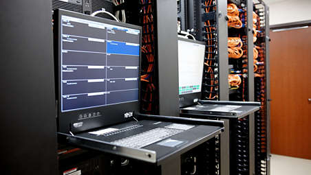
Building a virtual network with open, scalable solutions
Connect your teams with a scalable solution that enables easy sharing of patient-centric histology data across organizations and between sites. Build virtual networks across labs by adding new locations and users. IMS offers scalable, tiered server and storage options that grow with you. You can choose from three different storage plans to suit your needs. The open platform empowers new interoperabilityoptions as your needs and priorities change. The system easily interfaces with multiple LIS, a broad array of hardware, third-party scanner vendors and image analysis software. - Streamlining digital workflow to enhance user experience
-
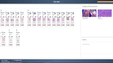
Streamlining digital workflow to enhance user experience
IMS offers specialized tools for measurement, annotation, collaboration, and archive management. Review cases quickly using fast slide-to-slide transitions and confer on cases using single-click collaboration connection. Case details are presented with a digital slide tray, which organizes the slides like an analog slide tray. It includes case-related documents, grossing images and case-specific notes. This overview of slides can indicate which slides have been viewed, annotated and if additional slides have been ordered. Access role-based, user-specific case lists along with related patient data and notes. - Enhancing tissue analysis with a dedicated slide viewer
-
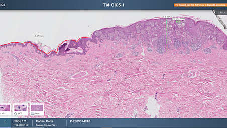
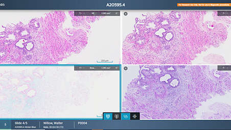
Enhancing tissue analysis with a dedicated slide viewer
Our dedicated slide viewer includes a rich set of tools for making annotations and accurate measurements, allowing pathologist to easily navigate tissue. Create a gallery of highlighted tissue features you can return to with the click of a button. Tag slides for follow-up in secondary workflows such as tumor board or panel discussions. Smart workflow algorithms include automatic image alignment, tissue detection, tissue presentation and single-click navigation. Shortcut keys help improve workflow and enhance productivity. - Enhancing real-time collaboration and case sharing
-
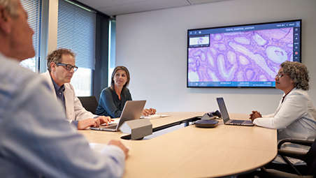
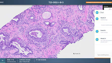
Enhancing real-time collaboration and case sharing
Connect with your colleagues anywhere in the world with our real-time collaboration tool. Share your slide viewing simultaneously in a manner that mimics a multi-headed microscope. Participants share control of the screen including annotation and measurement tools allowing them to collaborate on case analysis. - Flexible storage for digital pathology images
-
Flexible storage for digital pathology images
Pathology image management can be tailored to meet diverse needs—whether through a customizable on-premise storage configuration or in combination with secure cloud-based archiving. The Philips IntelliSite Pathology Solution offers robust repository management with LIS interoperability, network flexibility, and integration with existing systems. Cloud Data Services, for example in collaboration with our partner AWS, facilitates scalable, cost-effective storage with instant access, long-term retention, and a foundation for AI-powered integrated diagnostics.** - Improving the user experience and productivity
-
Improving the user experience and productivity
The Image Management System is your gateway to manage, review and analyze digitized slides. This next-generation software includes tools to optimize your daily caseload management, cases and images navigation and collaboration. Access whole slide images (WSI) in the feature-rich viewer in a streamlined, accessible diagnostic workflow. - Managing your workday just became easier
-
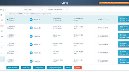
Managing your workday just became easier
IMS makes it possible to easily organize, review, manage and present digital cases. Facilitate organized personal worklists that are arranged and sorted according to your preference. Easily distinguish cases by status and case-related information such as patient name, number of slides per case and tissue type. Find the right case quickly with advanced search and filter functionality. - Adding the power of AI
-
Adding the power of AI
Philips IntelliSite Pathology Solution is designed as an open platform that can safely and securely share imaging and other relevant data to provide interoperability with third-party AI applications*
Integrating bi-directionally between LIS-IMS

Integrating bi-directionally between LIS-IMS
Integrating bi-directionally between LIS-IMS
Building a virtual network with open, scalable solutions
Building a virtual network with open, scalable solutions
Building a virtual network with open, scalable solutions
Streamlining digital workflow to enhance user experience
Streamlining digital workflow to enhance user experience
Streamlining digital workflow to enhance user experience
Enhancing tissue analysis with a dedicated slide viewer
Enhancing tissue analysis with a dedicated slide viewer
Enhancing tissue analysis with a dedicated slide viewer
Enhancing real-time collaboration and case sharing
Enhancing real-time collaboration and case sharing
Enhancing real-time collaboration and case sharing
Flexible storage for digital pathology images
Flexible storage for digital pathology images
Flexible storage for digital pathology images
Improving the user experience and productivity
Improving the user experience and productivity
Improving the user experience and productivity
Managing your workday just became easier
Managing your workday just became easier
Managing your workday just became easier
Adding the power of AI

Adding the power of AI
Adding the power of AI
- Integrating bi-directionally between LIS-IMS
- Building a virtual network with open, scalable solutions
- Streamlining digital workflow to enhance user experience
- Enhancing tissue analysis with a dedicated slide viewer
- Integrating bi-directionally between LIS-IMS
-
Integrating bi-directionally between LIS-IMS
Unify patient data in a streamlined workflow with bi-directional LIS-IMS integration. The updated viewer enables case review, access to multidisciplinary patient data and collaboration. Launch the IMS from the LIS worklist or synchronization of case-related clinical data between systems can be realized. - Building a virtual network with open, scalable solutions
-
Building a virtual network with open, scalable solutions
Connect your teams with a scalable solution that enables easy sharing of patient-centric histology data across organizations and between sites. Build virtual networks across labs by adding new locations and users. IMS offers scalable, tiered server and storage options that grow with you. You can choose from three different storage plans to suit your needs. The open platform empowers new interoperabilityoptions as your needs and priorities change. The system easily interfaces with multiple LIS, a broad array of hardware, third-party scanner vendors and image analysis software. - Streamlining digital workflow to enhance user experience
-
Streamlining digital workflow to enhance user experience
IMS offers specialized tools for measurement, annotation, collaboration, and archive management. Review cases quickly using fast slide-to-slide transitions and confer on cases using single-click collaboration connection. Case details are presented with a digital slide tray, which organizes the slides like an analog slide tray. It includes case-related documents, grossing images and case-specific notes. This overview of slides can indicate which slides have been viewed, annotated and if additional slides have been ordered. Access role-based, user-specific case lists along with related patient data and notes. - Enhancing tissue analysis with a dedicated slide viewer
-
Enhancing tissue analysis with a dedicated slide viewer
Our dedicated slide viewer includes a rich set of tools for making annotations and accurate measurements, allowing pathologist to easily navigate tissue. Create a gallery of highlighted tissue features you can return to with the click of a button. Tag slides for follow-up in secondary workflows such as tumor board or panel discussions. Smart workflow algorithms include automatic image alignment, tissue detection, tissue presentation and single-click navigation. Shortcut keys help improve workflow and enhance productivity. - Enhancing real-time collaboration and case sharing
-
Enhancing real-time collaboration and case sharing
Connect with your colleagues anywhere in the world with our real-time collaboration tool. Share your slide viewing simultaneously in a manner that mimics a multi-headed microscope. Participants share control of the screen including annotation and measurement tools allowing them to collaborate on case analysis. - Flexible storage for digital pathology images
-
Flexible storage for digital pathology images
Pathology image management can be tailored to meet diverse needs—whether through a customizable on-premise storage configuration or in combination with secure cloud-based archiving. The Philips IntelliSite Pathology Solution offers robust repository management with LIS interoperability, network flexibility, and integration with existing systems. Cloud Data Services, for example in collaboration with our partner AWS, facilitates scalable, cost-effective storage with instant access, long-term retention, and a foundation for AI-powered integrated diagnostics.** - Improving the user experience and productivity
-
Improving the user experience and productivity
The Image Management System is your gateway to manage, review and analyze digitized slides. This next-generation software includes tools to optimize your daily caseload management, cases and images navigation and collaboration. Access whole slide images (WSI) in the feature-rich viewer in a streamlined, accessible diagnostic workflow. - Managing your workday just became easier
-
Managing your workday just became easier
IMS makes it possible to easily organize, review, manage and present digital cases. Facilitate organized personal worklists that are arranged and sorted according to your preference. Easily distinguish cases by status and case-related information such as patient name, number of slides per case and tissue type. Find the right case quickly with advanced search and filter functionality. - Adding the power of AI
-
Adding the power of AI
Philips IntelliSite Pathology Solution is designed as an open platform that can safely and securely share imaging and other relevant data to provide interoperability with third-party AI applications*
Integrating bi-directionally between LIS-IMS
Integrating bi-directionally between LIS-IMS
Integrating bi-directionally between LIS-IMS
Building a virtual network with open, scalable solutions
Building a virtual network with open, scalable solutions
Building a virtual network with open, scalable solutions
Streamlining digital workflow to enhance user experience
Streamlining digital workflow to enhance user experience
Streamlining digital workflow to enhance user experience
Enhancing tissue analysis with a dedicated slide viewer
Enhancing tissue analysis with a dedicated slide viewer
Enhancing tissue analysis with a dedicated slide viewer
Enhancing real-time collaboration and case sharing
Enhancing real-time collaboration and case sharing
Enhancing real-time collaboration and case sharing
Flexible storage for digital pathology images
Flexible storage for digital pathology images
Flexible storage for digital pathology images
Improving the user experience and productivity
Improving the user experience and productivity
Improving the user experience and productivity
Managing your workday just became easier
Managing your workday just became easier
Managing your workday just became easier
Adding the power of AI
Adding the power of AI
Adding the power of AI
Technische specificaties
- IMS application server and storage
-
IMS application server and storage CPU - Dual socket Intel 6-core @ 2.3 GHz
RAM - 32GB
Storage - Protection against a single disk failure, e.g. RAID configuration or data replication.
- For usage with 5 scanners or more: Flash Disks.
Network connectivity for hospital - for hospital: 1 x 1 GB/s Ethernet
Network connectivity to scanner(s) - 1 x 1 GB/s Ethernet for up to 2 scanners
- 1 x 10 GB/s for 3 or more scanners
- 1 x 10 GB/s for 1 scanner with raw output
- to archiving system (optional): 1 x 10 GB/s Ethernet
- to network share (optional): 1 x 10 GB/s Ethernet
-
- Minimum requirements of client computer to run the IMS Viewer Software
-
Minimum requirements of client computer to run the IMS Viewer Software CPU - Minimum: Intel Xeon E5-1620 v3 @ 3.50 GHz or similar
RAM - Minimum: 8 GB physical memory
Connectivity - Minimum: 100 Mbit/s Ethernet
Video card - Minumum: GPU Memory: 4 GB, GeForce GTX 1050 Ti or similar, Memory bandwith: 112 GB/s
-
- Software requirements on client computer
-
Software requirements on client computer Operating system - Microsoft Windows 8, 8.1 or 10-64 bit
Browser software supporting HTML 5 standard - Recommended browsers: Chrome or Chromium-based Microsoft Edge
- Other supported browser: Internet Explorer
Other software - PDF Viewer (for viewing PDF documents, e.g. reports manually uploaded to cases)
- Email client (for correct functioning of the email link button)
-
- IMS application server and storage
-
IMS application server and storage CPU - Dual socket Intel 6-core @ 2.3 GHz
RAM - 32GB
-
- Minimum requirements of client computer to run the IMS Viewer Software
-
Minimum requirements of client computer to run the IMS Viewer Software CPU - Minimum: Intel Xeon E5-1620 v3 @ 3.50 GHz or similar
RAM - Minimum: 8 GB physical memory
-
- IMS application server and storage
-
IMS application server and storage CPU - Dual socket Intel 6-core @ 2.3 GHz
RAM - 32GB
Storage - Protection against a single disk failure, e.g. RAID configuration or data replication.
- For usage with 5 scanners or more: Flash Disks.
Network connectivity for hospital - for hospital: 1 x 1 GB/s Ethernet
Network connectivity to scanner(s) - 1 x 1 GB/s Ethernet for up to 2 scanners
- 1 x 10 GB/s for 3 or more scanners
- 1 x 10 GB/s for 1 scanner with raw output
- to archiving system (optional): 1 x 10 GB/s Ethernet
- to network share (optional): 1 x 10 GB/s Ethernet
-
- Minimum requirements of client computer to run the IMS Viewer Software
-
Minimum requirements of client computer to run the IMS Viewer Software CPU - Minimum: Intel Xeon E5-1620 v3 @ 3.50 GHz or similar
RAM - Minimum: 8 GB physical memory
Connectivity - Minimum: 100 Mbit/s Ethernet
Video card - Minumum: GPU Memory: 4 GB, GeForce GTX 1050 Ti or similar, Memory bandwith: 112 GB/s
-
- Software requirements on client computer
-
Software requirements on client computer Operating system - Microsoft Windows 8, 8.1 or 10-64 bit
Browser software supporting HTML 5 standard - Recommended browsers: Chrome or Chromium-based Microsoft Edge
- Other supported browser: Internet Explorer
Other software - PDF Viewer (for viewing PDF documents, e.g. reports manually uploaded to cases)
- Email client (for correct functioning of the email link button)
-
Related products
Alternative products
-
Pathology Scanner SG(i)60
- Operational excellence by scanning small batches in parallel.
Product bekijken
-
Pathology Scanner SG300
- Premium scanner that delivers best in class digitization for large labs.
Product bekijken


-
Pathology Scanner SG(i)60
SG60 is designed to accommodate laboratories with a lean workflow and need to scan small batches of slides to achieve operational excellence and short turnaround times by scanning batches in parallel. With a high throughput, high first time right rate and load and walk away scanning, the SG60 enables you to digitize your histology samples and obtain high quality clinical diagnostic images for routine use and integrated pathology networks. Pathology Scanner SGi* with native configurable DICOM JPEG and JPEG XL output helps reduce file sizes without compromising diagnostic quality, unlocking significant storage savings and performance improvements.
Product bekijken
-
Pathology Scanner SG300
SG300 is designed to accommodate laboratories for high volume labs that want to maximize scanner utilization and further reduce the total cost of ownership per slide by means of overnight scanning. With a high throughput, high first time right rate and load and walk away scanning, the SG300 enables you to digitize your histology samples and obtain high quality clinical diagnostic images for routine use and integrated pathology networks. Pathology Scanner SGi* with native configurable DICOM JPEG and JPEG XL output helps reduce file sizes without compromising diagnostic quality, unlocking significant storage savings and performance improvements.
Product bekijken
- The Philips Digital Pathology solution is a bundle of: Pathology Scanner Second Generation • Image Management System Viewer • Image Management System Application Server and Storage Software • High quality 27” monitor validated with the IVD solution (US only)
- *PIPS enables iSyntax files, and with the Software Development Kit (SDK) third party companies can use this for AI capabilities.
- Philips IntelliSite Pathology Solution is in conformance with Regulation (EU) 201 7/746 of the European Parliament and of the Council of 5 April 2017 on In Vitro Diagnostics (IVDR).
- **Third party claims from AWS, which may not apply in all markets and may be updated from time to time. The functionalities and benefits of the solution depend on customer-specific configuration and use. Please contact your local Philips representative for (market) availability.
- The Software Development Kit (SDK) and File Viewer is not intended for diagnostic, monitoring or therapeutic purposes or in any other manner for regular medical practice.